


Research
Microscopy Core Facility
The College of Science and Mathematics (CSM) Microscopy Core (MC) provides access to confocal, deconvolution, super resolution and other optical microscope systems that are useful for multi-color imaging of live and fixed cells and tissue samples, and high-content screening.
This state-of-the-art microscopy facility serves Kennesaw State University (KSU) and external researchers by providing microscopy related expertise, training and assistance for advancing their projects on various model organisms. Additionally, MC offers software packages on high-end workstations for image processing and analysis.
New and current users are expected to read and sign the following User Guide: KSU MCF User Guide. DocuSign is required for KSU faculty and staff.
Equipment
-
Zeiss LSM 900 Confocal microscope
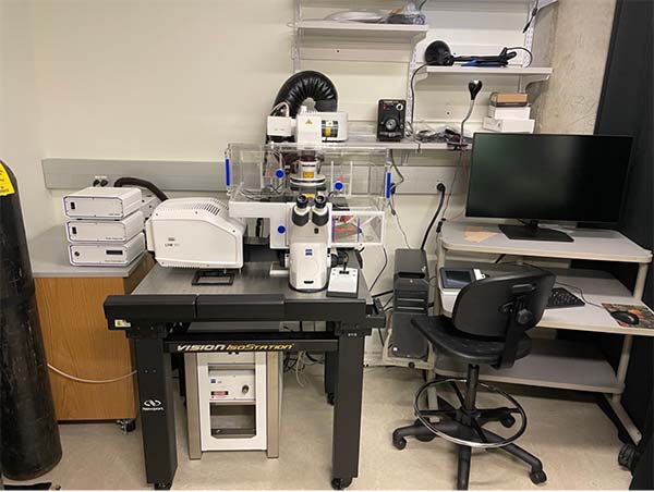
This is an inverted confocal microscope equipped with four solid-state lasers (405, 488, 555, and 639nm). The LSM 900 confocal is equipped with two GaSP PMT detection modules, enabling simultaneous four-beam acquisition. The microscope is equipped with an incubation chamber with controlled temperature and humidity for prolonged live cell imaging. A full range of objectives (e.g., 10x, 20x, 40x, 63x (oil immersion), 100x (oil immersion)) is installed on the microscope. This system is suitable for routine fixed and live imaging of either prepared specimens or live cells in conventional tissue culture vessels. Flow-through capabilities for long-term live imaging, including feeding kinetics, can be accommodated with this system. -
Leica TCS SP8 Tau-STED 3x Super-Resolution Microscope
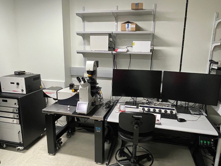
This is an inverted confocal microscope equipped with four solid-state lasers (405, 488, 555, and 639nm). The LSM 900 confocal is equipped with two GaSP PMT detection modules, enabling simultaneous four-beam acquisition. The microscope is equipped with an incubation chamber with controlled temperature and humidity for prolonged live cell imaging. A full range of objectives (e.g., 10x, 20x, 40x, 63x (oil immersion), 100x (oil immersion)) is installed on the microscope. This system is suitable for routine fixed and live imaging of either prepared specimens or live cells in conventional tissue culture vessels. Flow-through capabilities for long-term live imaging, including feeding kinetics, can be accommodated with this system. -
Zeiss Axio Imager M2 Upright Compound Microscope
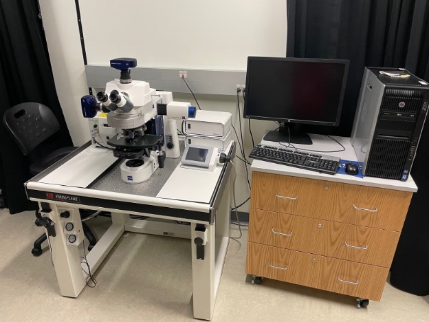
This is an upright compound microscope equipped with 4-color epifluorescence and an Apotome2 slider module. The Axio Imager is capable of conventional epifluorescence as well as phase illumination. Two cameras for high-resolution (4K compatible) imaging (mRM mono, mRM color) are mounted on the microscope for use. A full range of objectives (e.g., 5x, 10x, 20x, 40x, 63x (oil immersion) are installed on the microscope. Deconvolution capabilities are possible using the Apotome2 slider.
Fees and Scheduling
-
Fees
Please email the listed facility coordinator to inquire about the current usage rates. -
Scheduling
Users are limited to advance scheduling of one 2-hour block of time during core hours (defined as 9AM-5PM, Monday-Friday). Longer imaging runs can be accommodated after 5PM and/or on weekends. Users may schedule extra time on the day of use if available. Laboratories are asked to refrain from scheduling more than two 2-hour blocks in a single day on the same instrument.
Publications
In the acknowledgement section for publications resulting from work performed on the Confocal microscopes in the KSU Microscopy Core Facility, please provide the following statement:
“The authors would like to acknowledge Kennesaw State University Academic Affairs for support of the Microscopy Core Facility, which made possible the research necessary for the completion of in this project.”
If the work was performed on the Zeiss AxioImager M2 microscope, please provide the following statement:
“The authors would like to acknowledge Kennesaw State University Academic Affairs and Georgia BIO for support of the Microscopy Core Facility, which made possible the research necessary for the completion of in this project.”
RESERVE A MICROSCOPE
Kennesaw State University
Kennesaw Campus (maps)
Science Laboratory Building #105
Room SL 5049
105 Marietta Dr.
Kennesaw, GA 30144